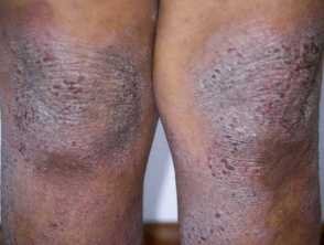
Revolutionary Imaging Technique for Atopic Dermatitis: Line-Field Confocal Optical Coherence Tomography (LC-OCT)
Recent advances in dermatological imaging have highlighted the potential of Line-field confocal optical coherence tomography (LC-OCT) for effectively visualizing inflammatory skin conditions. According to a promising study published in the Journal of the European Academy of Dermatology and Venereology, this innovative noninvasive technology offers rapid diagnostic capabilities, particularly for patients suffering from atopic dermatitis across various skin types.
Understanding LC-OCT and Its Benefits
What is LC-OCT?
LC-OCT combines the high cellular resolution of Reflectance Confocal Microscopy (RCM) with the impressive imaging depth of optical coherence tomography. This results in the creation of intricate 3D images that allow for detailed insights into skin health without invasive procedures.
How Does LC-OCT Differ from Other Imaging Techniques?
- Reflectance Confocal Microscopy (RCM): Provides high-resolution images parallel to the skin surface, primarily for superficial observation.
- Optical Coherence Tomography (OCT): Offers deeper imaging but lacks the high detail obtained from RCM.
LC-OCT addresses these limitations by capturing images in both horizontal and vertical planes, providing a more comprehensive view of skin conditions.
Study Overview: Efficacy of LC-OCT in Atopic Dermatitis
Research Details
Conducted at the Rutgers Center for Dermatology, the study included ten patients diagnosed with clinically evident atopic dermatitis. Researchers employed both LC-OCT and RCM to examine lesional and perilesional skin under a variety of conditions.
Key Methods
- Image Acquisition: Both horizontal and vertical images were captured from lesional and perilesional skin.
- Quantitative Analysis: Researchers assessed:
- Epidermal thickness
- Dermo-epidermal junction undulation
- Visual Analysis: Detailed microscopic features were analyzed, including:
- Spongiosis
- Exocytosis
- Perivascular inflammation
Significant Findings
- Epidermal Thickness: Atopic dermatitis lesions showed an increase in thickness compared to healthy skin, characterized by a thicker stratum corneum.
- Characteristic Features: LC-OCT successfully identified inflammatory markers linked to atopic dermatitis, paralleling the findings from RCM. Notably, some features were also found in clinically unaffected skin, indicating potential subclinical alterations.
Implications for Clinical Practice
Noninvasive Monitoring Tool
The insights gained from this study underscore LC-OCT’s potential as a fast and noninvasive tool for diagnosing and monitoring atopic dermatitis, an especially crucial asset for patients with darker skin types. Traditional assessment methods often rely on visual signs like redness, which can be misleading, especially in darker phototypes.
Quotes from Researchers
“This study supports the potential of LC-OCT for assessing inflammatory skin pathologies,” noted the study authors. “The ability to visualize microscopic features of atopic dermatitis lesions quickly and effectively is particularly important in improving patient outcomes.”
Conclusion: The Future of Dermatological Diagnostics
The study of LC-OCT marks a significant advancement in dermatological imaging. By offering a detailed snapshot of inflammatory conditions without invasive biopsies, this technology could revolutionize how dermatologists diagnose and monitor conditions like atopic dermatitis.
Further Reading
- Explore the potential applications of Reflectance Confocal Microscopy in treating dermatitis.
- Review comprehensive studies on various imaging technologies in dermatology.
With ongoing research and development, LC-OCT may soon become a standard in dermatological practice, enhancing the accuracy of diagnoses for millions suffering from skin conditions.


